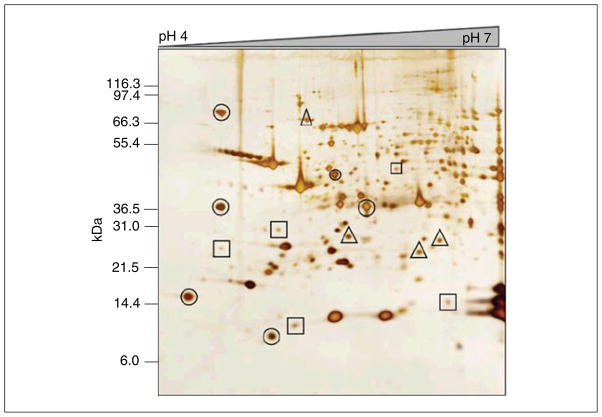
Figure 10.25.3.
200 μg of whole-tissue extract from mouse myocardium run on pH 4 to 7 two-dimensional gel and visualized with an MS-compatible silver stain. Protein spots that will likely provide reliable protein identification from a single gel (circled), as well as protein spots that will likely need to be pooled from multiple gels (squares), are indicated. Also shown are protein spots (triangles) that may or may not contain sufficient protein for identification, depending upon the MS instrumentation used.

