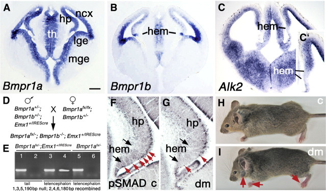
Figure 1.
Generating double mutant mice deficient in Bmpr1a and Bmpr1b. A–C, Coronal sections through the telencephalon of control mice at E11.5, processed with in situ hybridization to show expression of Bmpr1a (A), Bmpr1b (B), and a third type I BMP receptor gene, Alk2 (C). Bmpr1a is expressed throughout the telencephalic neuroepithelium including the cortical hem; Bmpr1b is expressed more selectively in the dorsal telencephalon with a hot spot of expression at the hem; Alk2 is expressed broadly in the dorsal and ventral telencephalon, but not in the cortical hem (C, C′). D, Breeding strategy used to generate mice lacking Bmpr1b constitutively and Bmpr1a conditionally, the Bmpr1afx/−;Bmpr1b−/−;Emx1+/IREScre double mutant genotype. E, PCR analysis of DNA extracted from the tail and telencephalon of E12.5 Bmpr1afx/−;Emx1+/IREScre and Bmpr1afx/− mice. Primers fx1 and fx4 (Mishina et al., 2002) amplified a 180 bp fragment from Bmpr1afx/−;Emx1+/IREScre telencephalon (lane 4), indicative of Cre-mediated recombination of Bmpr1afx. The 180 bp “recombined” band was not amplified from tail tissue (lane 2) or Bmpr1afx/− telencephalon (lane 6). Primers fx3 and fx5 amplified a 190 bp fragment from the Bmpr1a constitutive null allele in all three tissue samples (lanes 1, 3, 5). F, G, Coronal sections through the hem region at E12.5 in a control mouse (c) and a double mutant (dm), immunostained for pSmad1/5/8, transcription factors activated downstream of BMP signaling. The red arrows indicate pSmad1/5/8-IR cells in the control hem (F) but virtually no pSmad1/5/8-IR cells in the double mutant hem (G). Outside the hem, in the hippocampal primordium, pSmad1/5/8-IR cells are dense along the ventricular surface (F, G). H, I, A young adult control mouse (H) and littermate double mutant (I). The double mutant is slightly smaller than the control; the red arrows indicate truncated digits and partial loss of facial hair (see Results). Abbreviations: hem, Cortical hem; hp, hippocampus; lge, lateral ganglionic eminence; mge, medial ganglionic eminence; ncx, neocortex; th, thalamus. Scale bar: (in A) A–C, 200 μm; C′, 100 μm; F, G, 50 μm.

