
Figure 2.
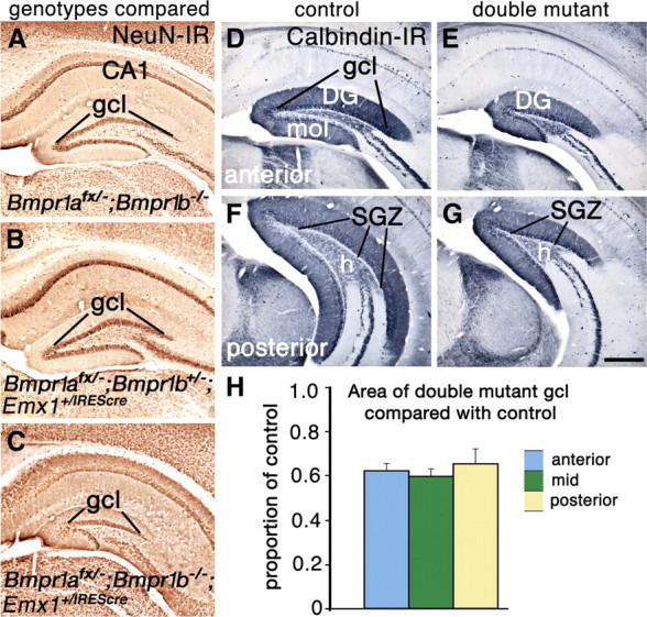
Loss of function of two BMP receptor genes leads to a reduced DG. A–G, Coronal sections through the hippocampus of 8-week-old (A–C) and 4-week-old (D–G) mice. A–C, Sections stained for NeuN immunoreactivity from mice lacking Bmpr1b and heterozygous for Bmpr1a (A), lacking Bmpr1a conditionally and heterozygous for Bmpr1b (B), or lacking both Bmpr1a and Bmpr1b (C). Only the double mutant shows a reduced gcl (C). D–G, Calbindin immunoreactivity reveals that the entire DG including the gcl, molecular layer (mol), hilus (h), and the SGZ is smaller in double mutants compared with controls. H, The cross-sectional area of the gcl in double mutants is ∼60% of that in control mice; this proportion is not significantly different at anterior, mid, or posterior levels of the DG. Data are represented as means ± SEM. Scale bar: (in G) A–G, 375 μm.