
Figure 4.
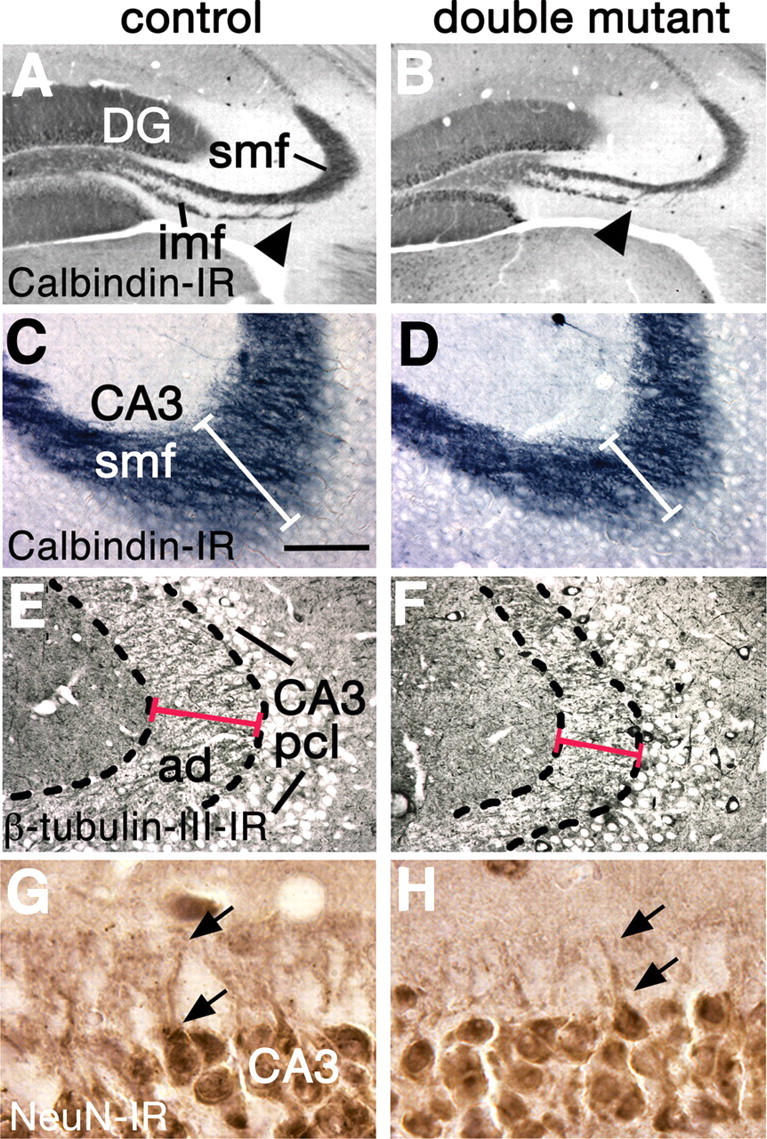
The projection from the DG to CA3 is abnormal in double mutants. A–H, Coronal sections through 8-week-old brains, processed for calbindin (A–D) class III β-tubulin (E, F) or NeuN immunoreactivity (G, H). A–D, In double mutant brains, the infrapyramidal mossy fiber bundle (imf) is truncated (A, B, arrowheads), and the intrapyramidal and suprapyramidal mossy fibers (grouped as smf) are reduced (A–D, white bars in C, D). E–H, The stratum lucidum of the hippocampus (E, F, broken outline) is filled with mossy fiber-recipient apical dendrites (ad) of CA3 neurons. CA3 apical dendrites are shorter in double mutants compared with controls (E, F, red lines); the arrows indicate the proximal parts of the apical dendrites of two neurons in G and H. Abbreviation: pcl, Pyramidal cell layer. Scale bar: (in C) A, B, 300 μm; C–F, 75 μm; G, H, 30 μm.