
Figure 6.
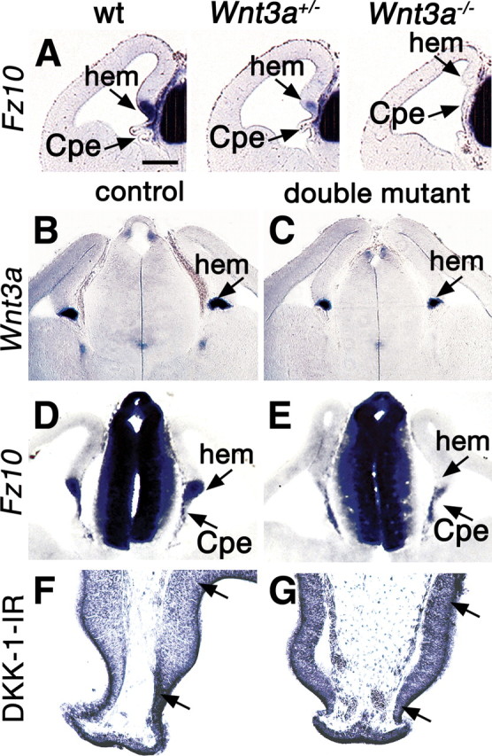
Defects of Wnt signaling in the cortical hem. A–G, Coronal sections through the E12.5 telencephalon processed for in situ hybridization. A, Expression of Fz10 in wild type (wt) and heterozygous and homozygous Wnt3a mutant brains. Fz10 expression fills the wild-type hem and appears in patches in the CPe; expression is weaker in the heterozygous Wnt3a mutant hem and absent from the homozygous mutant hem. B–E, The hem shows a smaller domain of Wnt3a expression in a double mutant compared with a control brain (B, C) and greatly reduced expression of Fz10 (D, E). F, G, A region (between arrows) including the hem and pDG shows immunoreactivity for a Wnt antagonist, DKK1. DKK1 immunoreactivity is more intense in this region in the double mutant (G) than in the control (F). Scale bar: (in A) A–E, 200 μm; F, G, 100 μm.