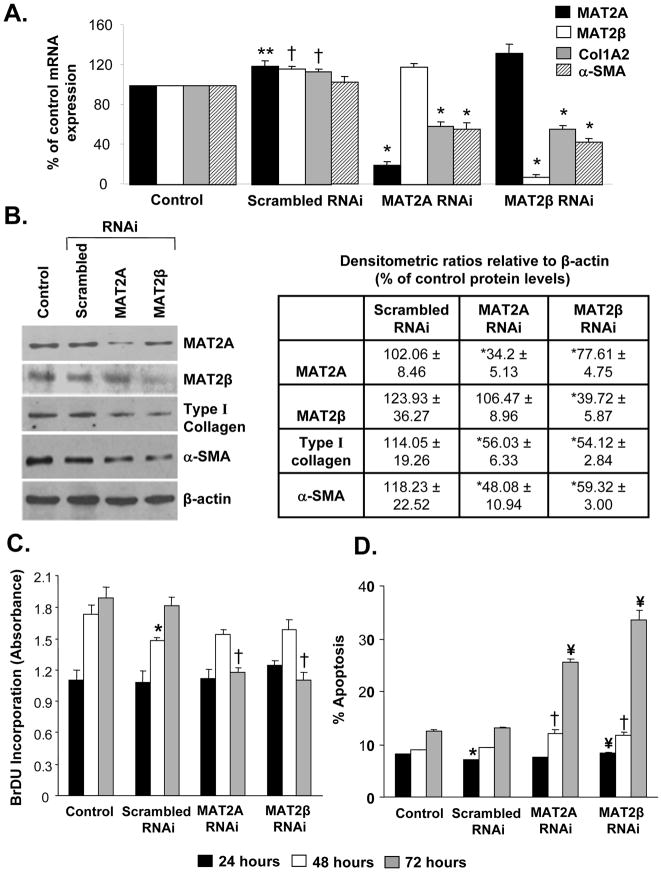
Fig. 4. Effect of MAT2A and MAT2β knockdown on activation, proliferation and apoptosis in human LX-2 cells.
Knockdown of MAT2A and MAT2β genes in LX-2 cells was performed as described in Methods. (A) Total RNA isolated from MAT2A or MAT2β knockdown cells was subjected to real-time RT-PCR analysis and expression of MAT2A, MAT2β , Col1A2 and α-SMA was compared to control or scrambled RNAi. Results represent Mean±SE from three experiments in duplicates for MAT2A, MAT2β , Col1A2 and four experiments for α-SMA; **p<0.05, †p<0.005 vs. control, *p<0.005 vs. scrambled RNAi. (B) Total cellular protein from MAT2A or MAT2β knockdown cells was subjected to Western blotting for detection of MAT2A, MAT2β, type I collagen and α-SMA and compared to control and scrambled RNAi. Representative images and densitometric analysis (Mean±SE) from three experiments is shown; *p<0.05 vs. scrambled RNAi. (C) Knockdown of MAT2A or MAT2β was done for 24, 48 or 72 hours and BrDU incorporation in RNAi-treated cells was compared to control or scrambled RNAi-treated cells. Results represent Mean±SE of three experiments in duplicates; *p<0.05 vs. control, †p<0.005 vs. scrambled RNAi. (D) Apoptotic cells in control or RNAi-treated samples were detected after 24, 48 or 72 hours using Hoechst staining as described in methods. Result represent Mean±SE of two experiments in duplicates; *p<0.005 vs. control, †p<0.05, ¥p<0.005 vs. scrambled RNAi.

