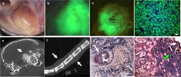
Figure 2.
Tumors in R-CreER+/−/SSM2+/− mice were detected in a variety of locations such as juxta-articular regions in limbs (a) that is a frequent location in human synovial sarcomas. The tumors showed fluorescence grossly (b and c) and in micrograph (d). Tumors in Tamoxifen exposed (b) and Tamoxifen unexposed (c) mice were fluorescent. CT imaging revealed calcifications (e, arrow) in some tumors such as the one shown in figure e located near the right orbit while others, such as the tail tumor showed in f (arrows show tumor masses), demonstrate exclusive soft tissue involvement.
Histology revealed a variety of tissue involvement such as bone (g, arrow) and subcutaneous tissue (h). White arrow in figure h show keratinized epithelial layer while green arrow show infiltrating tumor cells. All scale bars = 50 μm.

