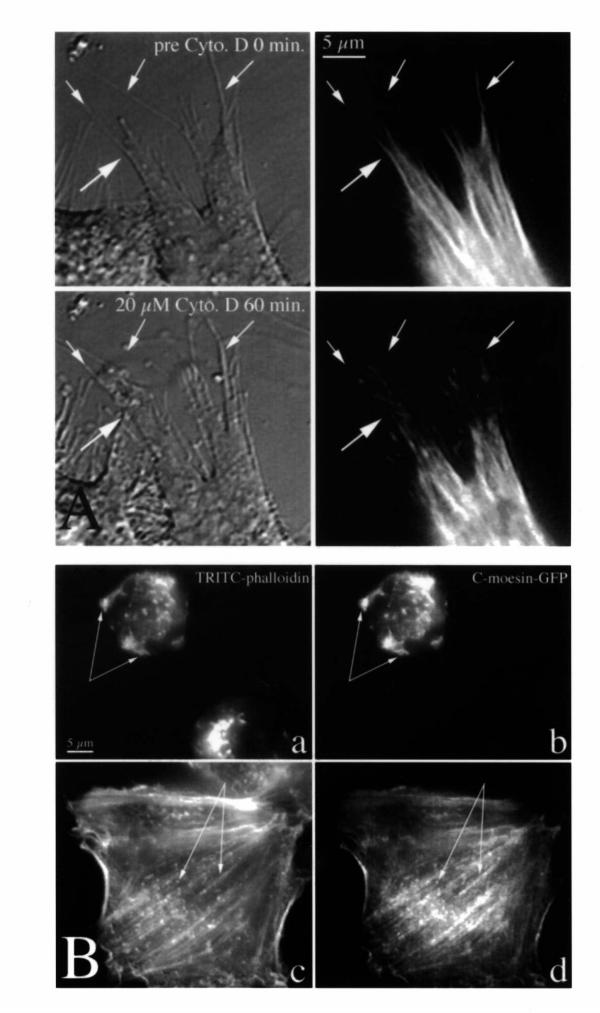
Figure 6.
C-moesin-GFP does not interfere with microfilament rearrangements during and after treatment with cytochalasin D. In (A), a transfected cell was imaged live by DIC (left) and C-moesin-GFP fluorescence (right) during treatment with cytochalasin D. The large arrow points at changes within one pseudopodium. The small arrows point to filopodia and retraction fibers. Note withdrawal and clumping of C-moesin-GFP fluorescence. In (B), cells were treated for 30 minutes with 20 μM cytochalasin D (a, b) and then fixed, or treated for 20 minutes and then allowed to recover for 1 hour after drug washout (c, d) before fixation. They were then stained with TRITC-phalloidin (a, c) and imaged for C-moesin-GFP fluorescence (b, d). Arrows point to identical spots in parallel images.

