
Figure 1. Vacuolar morphology is sensitive to growth conditions.
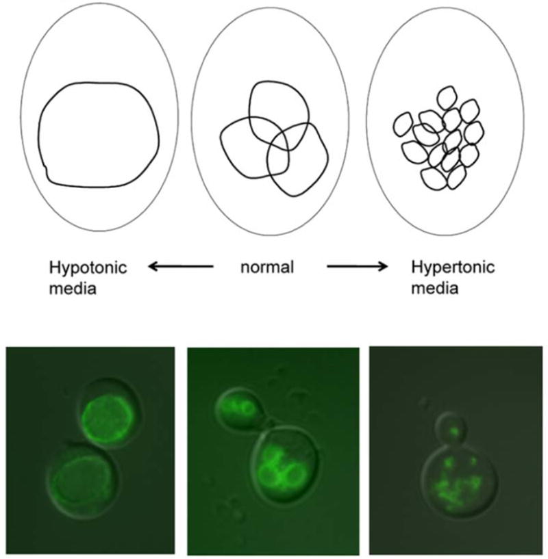
The vacuole can change in volume and vesicle number depending on extracellular condtions. Top: Under normal conditions, the cell has two to three medium-sized vacuoles. In hypo-osmotic media, a single large vacuole takes up most of the cell. Upon hyper-osmotic shock, the vacuole fragments into multiple small lobes. Bottom: Superimposed images of fluorescently stained vacuoles on yeast cells viewed under Nomarski optics. Vacuoles were visualized using a C-terminally GFP-tagged V-ATPase subunit (Vma2p-GFP) during log-phase growth under hypotonic conditions (synthetic complete medium), normal conditions (YEPD, buffered to pH 5 (100 mM added salt)), and hypertonic conditions (YEPD, buffered to pH 5 with 1 M NaCl added).