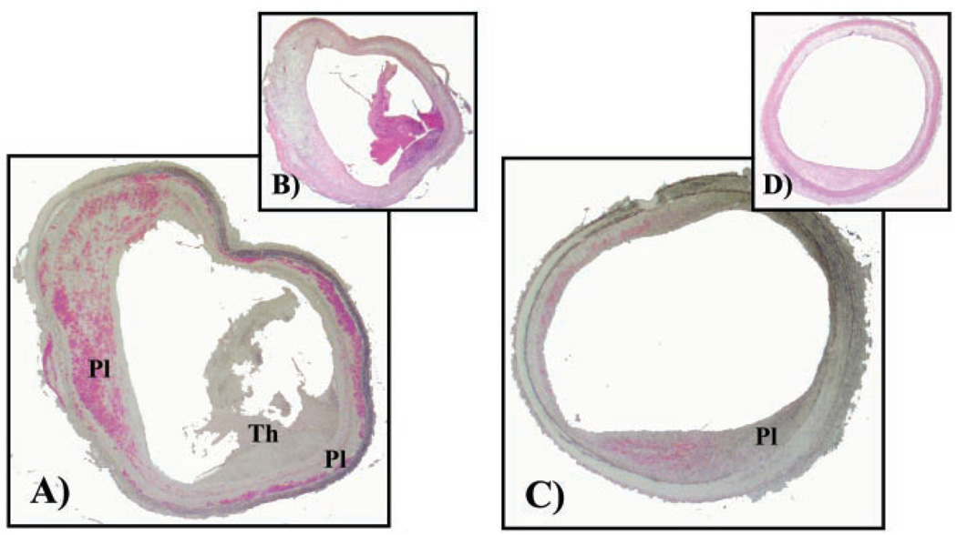
Figure 3.
Macrophage immunostaining (RAM1-11, DAKO Corp, fast red stain) and H&E staining of representative rabbit aortic cross section (5 µm) of placebo (A, B) and candesartan-treated (C, D) rabbits, respectively. Plaque area and thrombus are depicted by Pl and Th, respectively. Placebo sections(C, D) depict thrombus. Percentage area of macrophages was determined relative to total plaque area.

