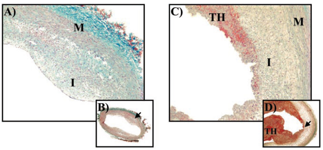
Figure 4.
Masson’s trichrome stain of aortic cross sections (5 µm) from candesartan-treated (A, B; high- [×10] and low- [×4] power, respectively) and placebo-control (C, D; high- and low-power, respectively) rabbits. Sections from placebo control rabbit show plaque erosion with overlying thrombus (TH). Blue staining denotes collagen. I indicates intima; M, media.

