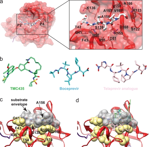
FIGURE 3.
a, amino acid residues of the substrate-binding pocket of NS3 protease. Residues capable of interacting with a peptide substrate are shown. Catalytic residues are underlined. b, examples of NS3 protease inhibitors. The macrocyclic TMC435 (Protein Data Bank code 3KEE) (99) and linear boceprevir (code 2OC8) (51) and telaprevir analog (code 2P59) (100) are shown. c, NS3 substrate envelope. The van der Waals surface of the NS3/NS4A junction peptide (gray) is shown relative to amino acids implicated in drug resistance (yellow). d, inhibitors extend beyond the substrate envelope (same as in c but showing the positions of the inhibitors). Inhibitor locations are based on crystal structures (51, 99, 100).

