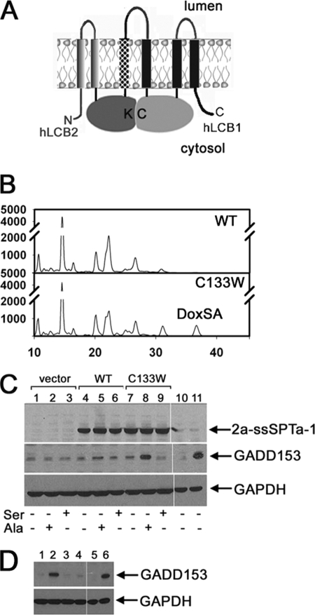
FIGURE 6.
1-deoxySa induces ER stress. A, model of the hLCB2-ssSPT-LCB1 triple fusion SPT in which ssSPT (stippled) is inserted between hLCB2 and hLCB1. Topologies are based on hydropathy analyses as well as preliminary experimental data as described in the text. Cys-133, which when mutated to tryptophan causes HSAN, is indicated (C). The pyridoxal phosphate-binding lysine in hLCB2, which in the crystal structure of S. paucimobilis lies directly across the dimer interface from Cys-133 (9), is also indicated (K). B, CHO-LyB cells expressing mutant human LCB2a-ssSPTa-LCB1C133W (bottom panel), but not wild-type LCB2-ssSPTa-LCB1 (top panel), fusion SPT accumulate 1-deoxySa (DoxSA). C, cells transfected with vector (lanes 1–3), or plasmids expressing hLCB2a-ssSPTa-LCB1 (WT; lanes 4–6), or hLCB2a-ssSPTa-LCB1C133W (C133W; lanes 7–9) were grown in Ham's F-12 with or without 10 mm serine or 10 mm alanine for 48 h. As controls, untransfected cells were treated with vehicle (lane 10) or 20 μg/ml tunicamycin for 5 h (lane 11). LCB2a-ssSPTa-LCB1, GADD153, and glyceraldehyde-3-phosphate dehydrogenase (GAPDH) were visualized by immunoblotting following fractionation of total cellular proteins by SDS-PAGE. D, untransfected CHO-LyB cells were treated with vehicle (0.4% bovine serum albumin/phosphate-buffered saline/ethanol) (lane 1), 1 μm 1-deoxySa (lane 2), 1 μm dihydrosphinganine (lane 3), 1 μm sphingosine (lane 4), 0.25% DMSO (lane 5), or 20 μg/ml tunicamycin in DMSO (lane 6). GADD153 and glyceraldehyde-3-phosphate dehydrogenase (GAPDH) were visualized by immunoblotting as above. Results are representative of data obtained in three independent experiments.

