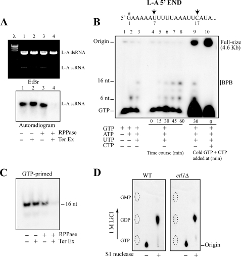
FIGURE 1.
GTP-primed L-A transcript has diphosphate at the 5′-end. A, 32P-labeled, full-sized L-A transcripts made in vitro by L-A virions were treated with Terminator 5′-exonuclease (Ter Ex) and/or RNA 5′-polyphosphatase (RPPase) as indicated below the panel and separated in an agarose gel. L-A single-stranded RNA (ssRNA) was visualized by ethidium bromide staining (upper panel) or by autoradiography (lower panel). λ, λ-HindIII fragments as size markers. B, upper panel shows the 5′-terminal region of the L-A single-stranded RNA transcript. The first U and C appear at positions 7 and 17, respectively (indicated by arrows). The asterisk indicates the position that can be labeled with [α-32P]GTP in an in vitro transcription reaction. The lower panel shows transcription products made in vitro by L-A virions. Transcripts were labeled with [α-32P]GTP, separated in a 15% acrylamide gel, and visualized by autoradiography. Lanes 1–3, L-A virions were incubated for 30 min in a transcription reaction from which ATP, UTP, and CTP (lane 1), or UTP and CTP (lane 2), or CTP (lane 3) was omitted. Lanes 4–8, L-A virions were incubated for the times indicated below the panel in a CTP-omitted transcription reaction. Lane 9, L-A virions were incubated in a CTP-omitted transcription reaction. After a 30-min incubation, excess amounts (1 mm each) of nonlabeled GTP and CTP were added to the mixture, and the reaction was carried out 30 min further. Lane 10, same as in lane 9, except that the nonlabeled GTP and CTP were added at 0 min. The reaction was carried out for 60 min. The mobility of full-sized, 16-nt, and 6-nt transcripts as well as unincorporated GTP is indicated. The 16-nt transcripts comigrated with bromphenol blue (BPB) in this gel system. C, [α-32P]GTP-primed 16-nt transcripts made by L-A virions in a CTP-omitted reaction were treated with RNA 5′-polyphosphatase and/or Terminator 5′-exonuclease as indicated below the panel and separated in a 15% acrylamide gel. An autoradiogram of the gel is shown. D, [α-32P]GTP-primed 16-nt transcripts made by L-A virions isolated from CTL1 (left panel, WT) or ctl1Δ (right panel, ctl1Δ) strain were purified in a 15% acrylamide gel. The transcripts were treated with or without S1 nuclease and analyzed on PEI cellulose with 1 m LiCl. The positions of nonlabeled GTP, GDP, and GMP are indicated by the dotted ovals.

