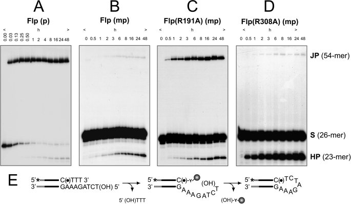
FIGURE 4.
Strand joining by Flp, Flp(R191A), and Flp(R308A) assayed using P half-site and MeP half-site substrates. The half-site substrates in the strand joining assays contained a free 5′-hydroxyl on the bottom (nonscissile) strand. A–D, reactions were analyzed by denaturing urea-PAGE to detect the products of strand joining (JP) and hydrolysis (HP). A and B, the reactions of wild type Flp with the P half-site (A) or the MeP half-site (B) are shown. C and D, only the MeP half-site reactions are shown for Flp(R191A) (C) and Flp(R308A) (D) because they were inactive on the P half-site (data not shown). The strand joining reaction mediated by the attack of the bottom strand 5′-OH on the P- or MeP-tyrosyl intermediate is schematically illustrated in E. The asterisk indicates the 32P label at the 5′-end of the top (scissile) strand, (●) denotes the scissile P or MeP, and (HO) represents the 5′-terminal hydroxyl group.

