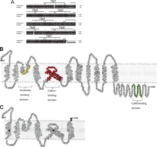
FIGURE 5.
Sequence analysis of AtNHXS1. A, comparison of deduced amino acid sequences of shuffled AtNHX1 and wild-type AtNHX1. The partial topology of the AtNHX1 protein, as proposed by protease protection assays (21) is shown above the sequence with open bars (predicted transmembrane segments). B, topological model of AtNHX1 based on Yamaguchi et al. (21). C, topological model of AtNHXS1; black residues indicate single amino acid substitutions.

