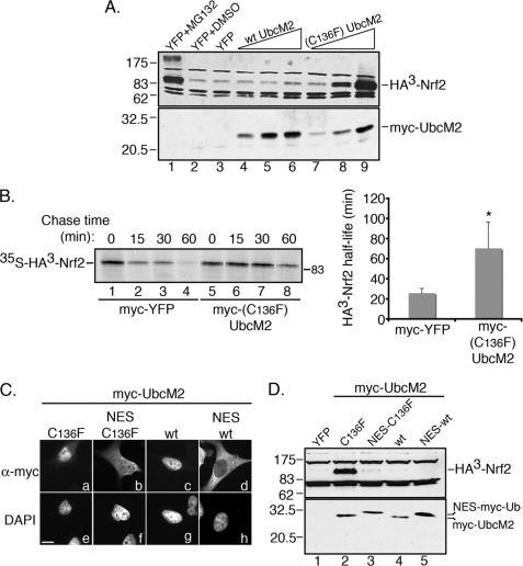
FIGURE 4.
(C136F)-UbcM2 increases the half-life of Nrf2 in the nucleus. A, co-transfection assay demonstrating dose-dependent stabilization of HA3-Nrf2 by (C136F)-UbcM2. MG132 treatment (lane 1) is a positive control for stabilization of the transcription factor and DMSO (lane 2) is the vehicle control for MG132. A plasmid expressing YFP was used to normalize the amount of transfected DNA (lanes 1–3). wt UbcM2 is expressed in lanes 4–6 and (C136F)-UbcM2 in lanes 7–9. HA3-Nrf2 was detected by α-HA (top panel) and Myc-UbcM2 proteins by α-UbcM2 (bottom panel). B, representative pulse-chase autoradiographic data used to determine the half-life of HA3-Nrf2 when co-expressed with either YFP (lanes 1–4) or (C136F)-UbcM2 (lanes 5–8). Chase times are indicated above the autoradiograph. The graph was obtained by averaging half-life measurements from three independent experiments. Error bars represent standard deviation, and the asterisk denotes statistical significance determined by two-tailed Student's t test (p < 0.0002). C, immunofluorescence analysis of the indicated Myc-UbcM2 proteins expressed in HeLa cells. Fusion of an NES relocalizes both (C136F)-UbcM2 and wt enzyme to the cytoplasm (compare panels a to b and c to d). Panels a–d are anti-Myc labeling and panels e–h are the corresponding DAPI images. The white bar represents 10 μm. D, same assay as A in HeLa cells testing how relocalizing (C136F)-UbcM2 to the cytoplasm influences Nrf2 stabilization. Experiments were performed at least three independent times.

