FIGURE 3.
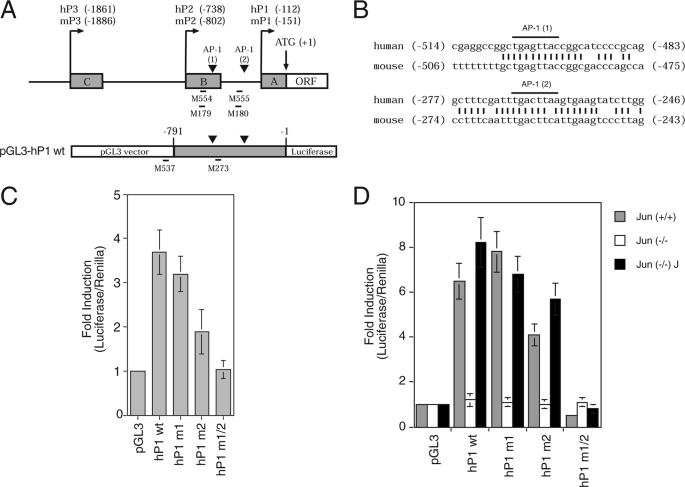
Two AP-1-binding sites contained in proximal Bcl-X promoter are required to respond to growth factor induction. A, schematic representation of the human and mouse Bcl-X promoter region covering the proximal promoters (P1, P2, and P3), and the alternative noncoding exons (boxes A, B, and C). The numbers shown refer to the distance upstream of the translation initiation codon (ATG). Untranslated exons are indicated in gray. The putative AP-1 sites are indicated by arrowheads. ORF, open reading frame. The genomic region containing the human P1 promoter (hP1), from nucleotides −791 to −1, was cloned upstream of the luciferase reporter gene in pGL3 plasmid; the construct obtained is indicated in the lower part of the panel. The locations and names of the oligonucleotides used in the ChIP assay are indicated. B, the AP-1 sites are conserved between mouse and human Bcl-X genes as both nucleotide sequences and positions into the genes. C, reporter constructs carrying the P1 promoter of the human Bcl-X gene as either wild type or mutated in the upstream AP-1, indicated as (1), downstream AP-1, indicated as (2), or at both sites (hP1wt, hP1 m1, hP1 m2, and hP1 m1/2) were cotransfected in HUVEC together with a constitutively expressing Renilla. Promoter activity values were normalized using Renilla activity, and the fold induction was calculated with respect to basal luciferase vector pGL3. D, the same constructs as in C were transfected in fibroblasts derived from c-Jun knock-out mouse (Jun (−/−)) and wild type control mouse (Jun (+/+)). As control, rescue of c-Jun was obtained by infection of knock-out fibroblasts with retrovirus for the overexpression of c-Jun (Jun (−/−) J). The mean values for three independent experiments ± S.D. of the mean are shown.

