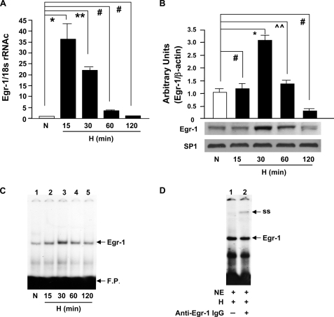
FIGURE 1.
Hypoxia-mediated induction of Egr-1 in THP-1 cells. THP-1 cells were exposed to hypoxia (H; 0.5% of oxygen) or normoxia (N) for the indicated times, and total RNA or nuclear extracts were isolated. A, real-time PCR analysis of Egr-1 expression was performed. Data are represented as the relative expression of mRNA for Egr-1 normalized to 18 S rRNA. B, immunoblotting with anti-Egr-1 IgG was performed on 5 μg/lane of nuclear protein from THP-1 cells, and results of multiple experiments were quantified. C, electrophoretic mobility gel shift assay was performed with 32P-labeled consensus Egr-1 oligonucleotide probe and nuclear extracts (3 μg/lane of nuclear protein) from THP-1 cells. D, supershift assay using anti-Egr-1 antibody was performed with 32P-labeled consensus Egr-1 oligonucleotide probe and nuclear extracts (3 μg/lane of nuclear protein) from THP-1 cells after 30 min of hypoxia. All experiments were repeated more than three times; representative bands are shown, and the mean ± S.E. (error bars) is shown. * (p < 0.0001), ** (p < 0.001), and ⋀⋀ (p < 0.05) indicate statistical significance; # indicates no statistical significance. F. P. indicates free probe. NE indicates nuclear extract.

