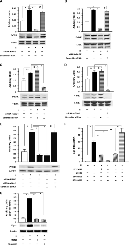
FIGURE 4.
Hypoxia-mediated activation of PKCβII, ERK1/2, and JNK leading to up-regulation of Egr-1 in THP-1 cells. Total protein, membrane protein, nuclear protein, and RNA were prepared from THP-1 cells exposed to hypoxia (H; 0.5% of oxygen) or normoxia (N) for the indicated times. A–D, immunoblotting with anti-phospho-ERK1/2 (P-ERK1/2) IgG (A and C) and anti-total-ERK1/2 (T-ERK1/2) IgG (A and C) or with anti-phospho-JNK (P-JNK) IgG (B and D) and anti-total-JNK (T-JNK) (B and D) was performed on 5 or 50 μg/lane of protein from THP-1 cells transfected with or without siRNA-RAGE (A and B) or siRNA-mDia-1 (C and D) and scramble siRNA and subjected to hypoxia for 10 min. E, immunoblotting with anti-PKCβII IgG was performed on 5 μg/lane of membrane protein from THP-1 cells transfected with or without siRNA-RAGE or siRNA-mDia-1 and scramble siRNA and subjected to hypoxia for 10 min. F and G, RNA and nuclear protein were prepared from THP-1 cells preincubated with or without inhibitors of PKCβ (LY379196, 30 nm), ERK1/2 (U0126, 5 μm), JNK (SP600125, 20 μm), and p38 (SB203580, 20 μm) and subjected to hypoxia for 15 (F) or 30 (G) min. F, real-time PCR analysis of Egr-1 expression was performed. Data are represented as the relative expression of mRNA for Egr-1 normalized to 18 S rRNA. G, immunoblotting with anti-Egr-1 IgG was performed on 5 μg/lane of nuclear protein from those THP-1 cells. All results of multiple experiments were quantified. * (p < 0.0001), ** (p < 0.001), and ⋀ (p < 0.05) indicate statistical significance; #, no statistical significance.

