Figure 1.

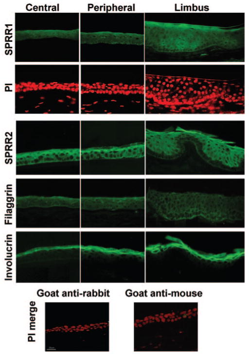
Immunofluorescence images of human cornea tissue sections showing reactivity against SPRR1, and -2, filaggrin, and involucrin. Bottom: Negative control images merged with images of propidium iodide counterstaining.

Immunofluorescence images of human cornea tissue sections showing reactivity against SPRR1, and -2, filaggrin, and involucrin. Bottom: Negative control images merged with images of propidium iodide counterstaining.