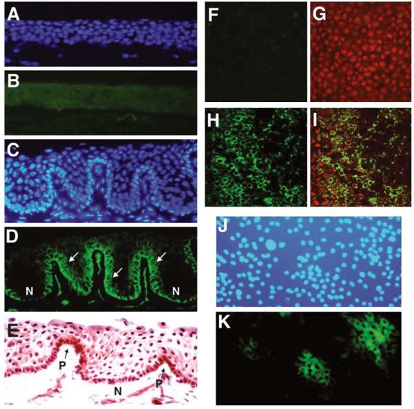
Figure 1.

Immunofluorescent (A–D, J, K) and immunohistochemical (E) stainings and laser scanning confocal microscopy (F–I) for ABCG2 transporter in human cornea (A, B, F, G), limbus (C–E, H, I), and primary cultured human limbal epithelial cells (J–K). (A, C, J): Hoechst 33342 counterstaining. (B): Cornea showing no staining for ABCG2. (D): Limbus showing brighter membrane staining for ABCG2 on certain basal cells (arrows), whereas other basal cells are negative (N). (E): Limbus showing cytoplasmic staining in some of basal cells (P). (K): Cultured limbal epithelial cells showing positive ABCG2 staining in clusters of small cells. Confocal microscopy showing ABCG2 stained on basal layer of whole-mounted human limbus (H, I) but not corneal basal layer (F, G). (G, I): Merged color images for ABCG2 (green) and propidium iodide (red). Original magnification × 400.
