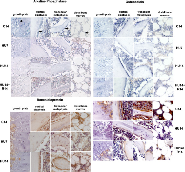
Figure 2.
Changes in the pattern of expression of genes involved in the osteogenic progression (AP, OC and BSP) induced by HU, and their reversibility. Gene expression analyzed by in situ hybridization for AP, OC and by immunohistochemistry (for OC and BSP). Control mice at 14 days (C14) and mice in HU for 7 (HU7) or 14 days (HU14) and for 14 days in HU, followed by 14 days of re-loading (HU14+R14). From left to right in each row: growth plate, cortical bone at mid-diaphysis, trabecular bone in the proximal region, bone marrow in the distal region of the tibia. Arrows in the first lane point to different cell types in these compartments, respectively pre-hypertrophic columnar chondrocytes, osteogenic cells of the endosteum, osteogenic cells lining trabeculae and bone marrow cells. Probes are described in M&M. Enlargement is 200×.

