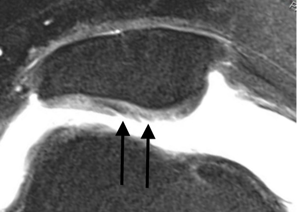
Figure 2.
Axial PD-weighted TSE MRI of a 29-year-old male with a recurrent LPD. MRI shows a fibrillation, fissuring, or erosion composing < 50% of the cartilage thickness at the central dome and the lateral facet of the retropatellar articular surface (black arrow). This finding is defined as a grade 2 disorder.

