Figure 5.
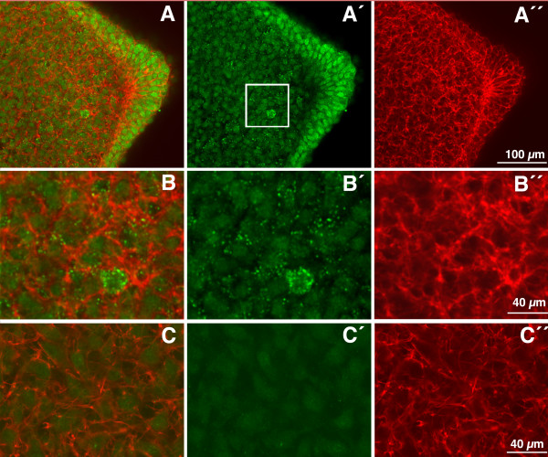
LTBP1 immunolabeling in the early limb bud. (A-B'') LTBP1 immunolabeling (green) counterstained with cytoplasmic phalloidin-TRITC labeling (red) of an early limb bud section (stage HH22) at the level of the AER. In all cases the merge images (A and B); the green channel for LTBP1 immunolabeling (A' and B'); and the red channel for actin labeling with phalloidin-TRITC (A'' and B'') are shown. (A-A") Note the strong labeling of the cells of the AER and the positive extracellular dotted labeling pattern in the underlying mesenchyme. (B-B'') detailed view of the region outlined by an square in A', showing the positive labeling of the matrix. Note the absence of overlapping between the cytoplasmic red labeling and the green spots indicative of its location in the pericellular space. (C-C'') Control section of a similar sample unexposed to the primary antibody for LTBP1.

