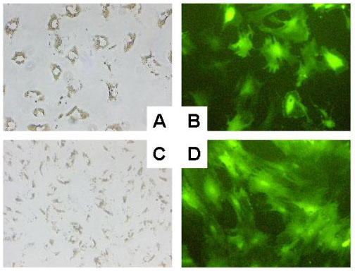
Figure 5.
Adenoviral infection of rHSCs and rMFBs with Ad5-CMV-EGFP. For infection of rHSCs/rMFBs with Ad5-CMV-EGFP 105 cells were seeded in 2 ml medium and infected 2 days later with 500 μl viral stock containing approximately 106 plaque forming units. Representative phase-contrast microscopy (A, C) and fluorescence microscopy (B, D) of rHSC (A, B) and rMFB (C, D) 48 hours after adenoviral infection are shown. Mock infected rHSCs or rMFBs are negative for EGFP-expression (not shown).

