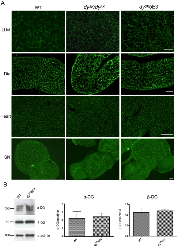
Figure 4. Normal expression of dystroglycans in dy3K/δE3 muscles.
(A) Cross-sections of limb muscle (Li M), diaphragm (Dia), heart and sciatic nerve (SN) from wild-type, dy3K/dy3K and dy3K/δE3 mice were stained with antibody IIH6 against α-dystroglycan. Bars, 50 µm. (B) Immunoblotting of glycoprotein preparations from wild-type and dy3K/δE3 skeletal muscle and quantitative measurement of α- and β-dystroglycan expression. Results are shown as means ± SEM. No significant difference in expression of α- and β-dystroglycan was noted between wild-type and dy3K/δE3 muscle (p = 0.8200 and p = 0.7527, respectively).

