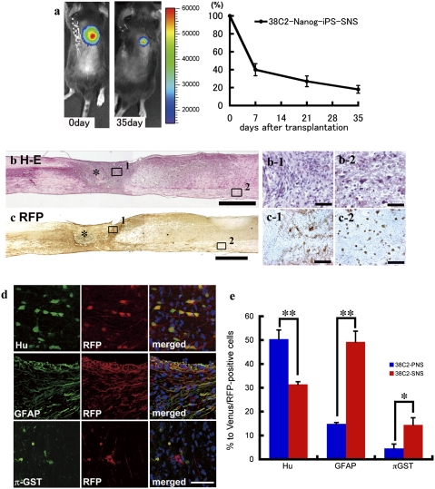
Fig. 2.
Transplanted SNSs derived from safe MEF-iPS clones survive without any evidence of tumorigenesis and differentiate into trilineage neural cells in the injured spinal cord. (A) Representative BLI images of a mouse in which CBRluc-expressing 38C2 iPS-SNSs were transplanted into the injured spinal cord (Left, immediately after transplantation; Right, 42 d after transplantation). Quantification of the photon intensity revealed that ≈60% of the grafted cells were lost within 7 d after transplantation, and ≈20% of the cells survived 35 d after transplantation. Values are means ± SEM (n = 6). (B) H&E and (C) anti-RFP DAB staining of sagittal sections of the spinal cord 42 d after injury (38C2 iPS-SNS transplanted). There was no evidence of tumorigenesis (B). No significant nuclear atypia was observed in magnified images of the boxed areas showing the lesion epicenter (B-1) or white matter caudal to the transplantation site (B-2). Grafted cells survived and were diffusely distributed rostral and caudal to the lesion site (C). Higher-magnification images of the boxed areas showing the lesion site (C-1) and white matter caudal to the lesion site (C-2). *Lesion epicenter. (D) Immunohistochemical analyses of 38C2 iPS-SNSs grafted into spinal cord 42 d after injury, revealing grafted cells double-positive for RFP and markers of neural lineages. (E) Quantitative analyses of Hu+ neurons, GFAP+ astrocytes, and π-GST+ oligodendrocytes. Values are means ± SEM (n = 3 each; *P < 0.05, **P < 0.01).

