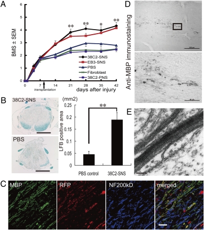
Fig. 3.
SNS derived from a safe MEF-iPS clone differentiate into mature oligodendrocytes and promote remyelination. (A) Time course of functional recovery of hind limbs evaluated by BMS. 38C2 iPS-SNS, n = 19; EB3 ES-SNS, n = 15; PBS, n = 12; adult fibroblasts, n = 13; 38C2 iPS-PNS, n = 13. *P < 0.05, **P < 0.01. (B) LFB staining of axial sections of the spinal cord at the lesion epicenter 42 d after injury; 38C2 iPS-SNS–transplanted (Upper Left) and PBS control (Lower Left) animals. Quantification of LFB-positive areas at the lesion epicenter 42 d after injury (Right, n = 7 each; **P < 0.01). (C) Immunohistochemistry of 38C2 iPS-SNS–derived mature oligodendrocytes (MBP+). Grafted cells were integrated into myelin sheath. (D) Anti-MBP DAB staining of sagittally sectioned spinal cord of a shiverer mouse 8 wk after transplantation. MBP+ myelin was detected in the area caudal to the lesion epicenter. (Lower) Higher-magnification image of the boxed area. (E) EM pictures of the injured spinal cord of a 38C2 iPS-SNS–grafted shiverer mouse exhibiting a prominent major dense line and intraperiod lines in multiple compacted lamellae. (Scale bars: B, 500 μm; D Upper, 200 μm; C and D Lower, 50 μm; and E, 0.1 μm.)

