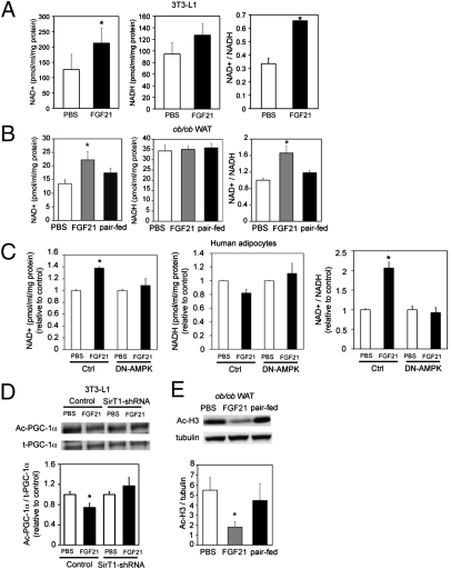
Fig. 2.
FGF21 increases cellular NAD+ and decreases H3 acetylation. (A) NAD+/NADH levels in 3T3-L1 adipocytes. 3T3-L1 adipocytes were treated for 3 d with FGF21 (4.0 μg/mL; black bars) or PBS (white bars). (B) NAD+/NADH levels in WAT from vehicle-treated (white bar), FGF21-treated (gray bar), and paired-fed (black bar) mice. n = 8 animals/group. (C) NAD+/NADH levels in human adipocytes transduced with adenovirus expressing control vector or DN-AMPK. Transduced adipocytes were treated for 3 d with FGF21 (4.0 μg/mL; black bars) or PBS (white bars). (D) Western blot and quantification of acetylated PGC-1α (Ac-PGC-1α) in 3T3-L1 adipocytes. 3T3-L1 adipocytes were transduced with adenovirus expressing SIRT1-shRNA and Flag–PGC-1α. Flag–PGC-1α was immunoprecipitated and blotted with pan acetylated lysine antibody. t-PGC-1α, total PGC-1α. (E) Western blot and quantification of acetylated H3 (Ac-H3) in WAT from vehicle-treated (white bar), FGF21-treated (gray bar), and paired-fed (black bar) mice. All data are averages of three independent experiments. n = 8 animals/group. *P < 0.05 (Student's t test).

