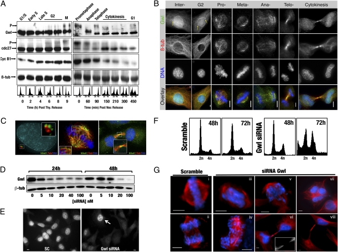
Fig. 1.
hGwl siRNA knockdown induces multiple mitotic defects. (A) HeLa cells were synchronized by either thymidine (Thy) or nocodazole (Noc) shake-off and released into fresh media. Samples were collected at the indicated times and processed for Western blot. Cell synchrony was confirmed by FACS. (B) HeLa cells were probed with anti-hGwl (green), anti-β-tubulin (red), and DAPI (DNA, blue). The maximum projections from 0.3-μm Z-sections are shown. Yellow arrows indicate the centrosomal localization of hGwl. (Scale bars, 5 μm.) (C) Enlargements of HeLa cells treated as per B. Cells were counterstained with γ- (interphase, red) or β-tubulin (mitotic, cytokinesis, red), hGwl (green), and DNA (blue). (D) Asynchronous HeLa cells were transfected with the indicated amounts of hGwl siRNA for 24 h and harvested immediately (24 h) or 1 d later (48 h). The levels of hGwl were analyzed by Western blot. (E) Asynchronous HeLa cells were transfected with 50 nM of siRNA and analyzed by immunofluorescence with anti-hGwl antibodies. Arrow indicates a nontransfected cell. (F) HeLa cells were treated as per D with either 50 nM of Scramble or hGwl siRNA and analyzed by FACS. (G) Asynchronous HeLa cells treated as in D were analyzed by immunofluorescence. For β-tubulin (red) and DAPI (DNA, blue). (Scale bars, 5 μm.)

