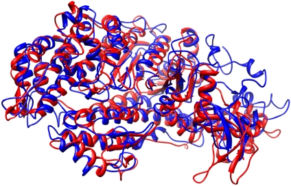
Fig. 3.
Comparison of the converged structure of myosin II in blue and myosin V (chain A of 1W8J) in red shown as secondary structure cartoons. The actin binding cleft in myosin II has closed during the refinement and the β-sheet has become more twisted. Generated with Chimera (11).

