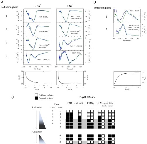
Fig. 4.
(A) Reduction of the NqrB-D346A mutant by 250 μM NADH in the absence and presence of 100 mM NaCl. (B) Kinetic phases of the oxidation of the NqrB-D346A mutant. In A and B blue lines show the difference spectra of the reduction steps associated with each transition, obtained by data fitting. Green lines show the difference spectra of the assigned redox steps using wild-type Na+-NQR as reported before (14). Bottom panels: Time course at the absorbance maximum (450 nm) of Na+-NQR (C) Scheme showing the redox transitions for the reduction and oxidation kinetics in the NqrB-D346A mutant. Open squares represent the oxidized state of the cofactor, and black squares the reduced form. The phases highlighted in gray show the transitions slowed down in the mutant enzyme.

