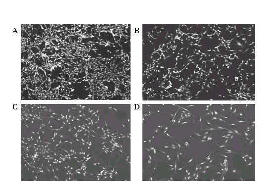
Figure 2.
Morphology of passage 6 BPAEC treated with the following concentrations of resveratrol: A) 0 μM; B) 10 μM; C) 25 μM; D) 50 μM. Cells were stained with rhodamine-phalloidin and viewed under 10X confocal microscopy. The same experiment was repeated three times, using passages 6-7 cells from two different preparation of BPAEC.

