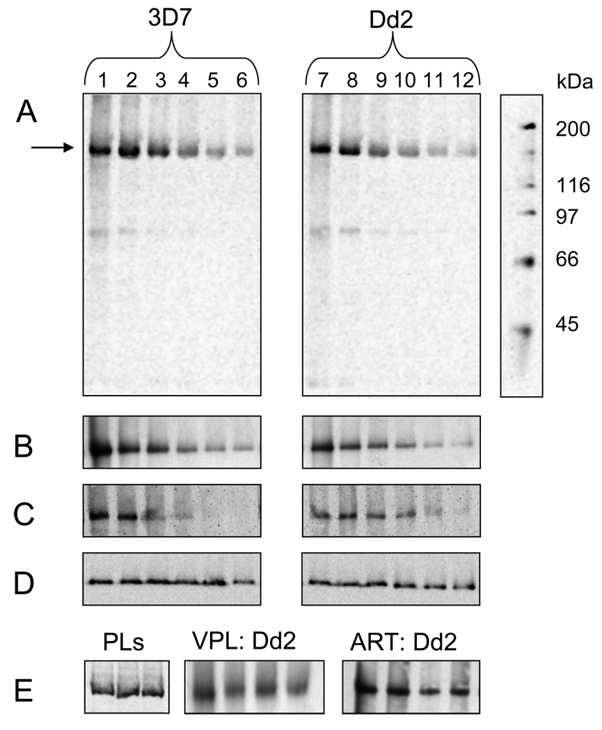
Fig. 1.
Characterization of AzBCQ photolabeling to partially purified 3D7 and Dd2 PfMDR1 isoforms reconstituted into proteoliposomes (PLs). Reaction conditions were as previously reported for PfCRT (13) with a few modifications. Briefly, equivalent amounts of purified PLs were diluted into 0.050 M Mes-Tris buffer at pH 5.2 and aliquots distributed to individual wells of a 96 well plate. A probe to protein molar ratio of approximately 100:1 was utilized in each experiment. 2.5 nmoles (50 µM final concentration) AzBCQ were added, the mixture was incubated at 37°C for 10 min, and then reacted under 254 nM UV light for 10 min. UV illumination was from a Spectroline 8W 60Hz bulb emitting 500 µW / cm2 that was positioned approximately 10 cm above the mixture. Photolabeling was quenched by the addition of 1X Laemmli buffer, and samples were incubated again at 37°C for 10 min before loading half the sample onto one of two 7.5% acrylamide Tris-HCl gels. Gels were run at 110V for 100 min., and immediately transferred to PVDF membrane for 16h at 40 mA and 4°C. Membranes were probed either with Anti-PentaHis-HRP (Qiagen) to detect PfMDR1 protein or Streptavidin-HRP (Amersham) to detect bound AzBCQ (see [13]). A. Representative streptavidin-HRP detection of AzBCQ photolabeling of 3D7 (left) and Dd2 (right) PfMDR1 PLs vs. competition with unlabelled CQ: lanes 1 & 7; no CQ competitor; lanes 2 – 6 and 8 – 12; 16-, 32-, 48-, 64- and 80-fold molar excess (800 µM, 1.6 mM, 2.4 mM, 3.2 mM, and 4.0 mM final concentration) of CQ relative to AzBCQ respectively. B. Representative streptavidin-HRP detection of AzBCQ photolabeling of 3D7 (left) and Dd2 (right) PfMDR1 PLs vs. QN competition: lanes 1 & 7- no QN; lanes 2 – 6 and 8 – 12; 5-, 10-, 20-, 40- and 80-fold excess of QN (250 µM, 500 µM, 1 mM, 2 mM and 4 mM final concentration) relative to AzBCQ, respectively. C. Representative streptavidin-HRP detection of AzBCQ photolabeling of 3D7 (left) and Dd2 (right) PfMDR1 PLs vs MQ competition: lanes 1 & 7- no MQ; lanes 2–6 and 8–12; 0.5-, 1-, 2.5-, 5- and 10-fold excess of MQ (25 µM, 50µM, 125 µM, 250 µM and 500 µM final concentration) relative to AzBCQ respectively. D. Representative PentaHis-HRP detection of 3D7 (left) and Dd2 (right) PfMDR1 PLs used in the CQ drug competition assay (Fig. 1A) showing approximately equal amounts of each protein isoform in all lanes. E, left. PentaHis HRP detection of 3 representative PL preparations. Equal volumes of 3D7 (lane 1), Dd2 (lane 2) and 7G8 (lane 3) PfMDR1 PLs show approximately equal protein:lipid ratio during reconstitution. E, center. Streptavidin-HRP detection of AzBCQ photolabeling of Dd2 PfMDR1 PLs with verapamil (VPL) competition. Lane 1 no VPL; lanes 2–4, 20-, 40- and 80-fold molar excess of VPL (1 mM, 2 mM, and 4 mM final concentration) relative to AzBCQ respectively. E, right. Streptavidin-HRP detection of AzBCQ photolabeling of Dd2 PfMDR1 PLs with artemisinin (ART) competition. Lane 1 no ART; lanes 2–4, 20-, 40- and 80-fold excesses of ART (1 mM, 2 mM, and 4 mM final concentration) relative to AzBCQ respectively.

