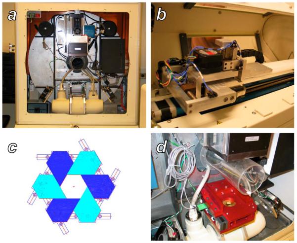
Figure 1.
The GE RS120 microCT and the additional components necessary for small animal radiotherapy. A: A view of the scanner gantry after removing the x-ray shield and animal stage housing. The large silver disk is the rotating gantry, with the x-ray tube, H-shaped collimator mounting bar, subject bore, and detector arranged bottom to top, respectively. B: The custom two-dimensional translation stage for subject positioning, mounted on top of the existing z-axis translation stage of the scanner. C: Schematic representation of a single stage of the variable-aperture collimator, formed by six sliding blocks mounted on linear tracks. D: The final two-stage collimator apparatus after installation on the H mounting bar of the scanner gantry.

