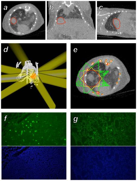
Figure 4.
Treatment of a murine spontaneous lung tumor with the microCT radiotherapy system. A-C: A MYC-induced tumor growing in the base of the right lung of a mouse was imaged with microCT, shown in axial, coronal, and sagittal sections with the tumor volume outlined in red. D: A treatment consisting of 8 beams of diameter 8 mm at isocenter with angular spacing of 45° was constructed in RT_Image to irradiate the target (red) to a dose of 2 Gy. E: Monte Carlo simulation of the dose delivered by this plan, with the 1, 1.4, and 1.8 Gy isodose contours shown in green, yellow, and red, respectively. F: γH2AX (green) and DAPI immunohistochemical sections from the target tumor. G: Corresponding immunohistochemical sections from the left lung that received an average dose of 0.3 Gy.

