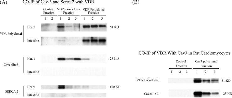
Figure 3.
A, Co-immunoprecipitation of Caveolin-3 and serca 2 with VDR antibodies. 600 ug of cardiomyocyte and intestinal protein lysates were incubated with VDR monoclonal and polyclonal antibody-coupled gel resins, and control gel resin respectively. Eluted fractions of IPed protein samples (20 ul each) were subjected to Western blot analysis. The blot was first probed with Caveolin-3 antibody (sc-5310) and then striped, and reprobed with Serca 2 antibody (sc-8094) and VDR antibody (sc-1008), respectively. B, Co-immunoprecipitation of VDR with caveolin-3 antibody. 600 ug of cardiomyocyte lysates were incubated with caveolin-3 antibody-coupled gel resin and control resin. Eluted fractions of immuno-precipitated samples (20 ul each) were subjected to Western blot analysis. The blot was first probed with caveolin-3 antibody (sc-5310) and then striped, and reprobed with VDR antibody (sc-1008).

