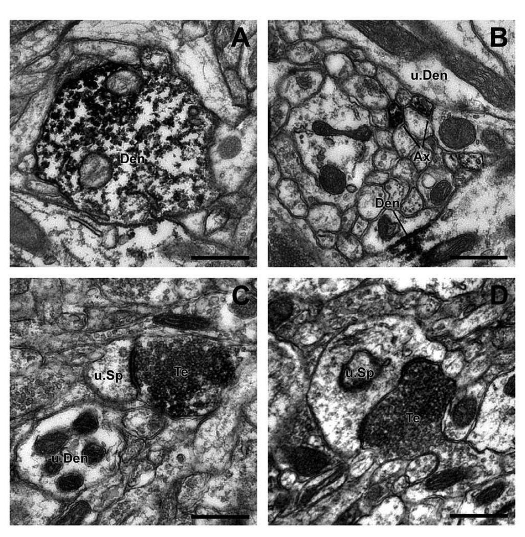
Figure 7.
Electron micrographs of Lrrk2-immunoreactive profiles in the monkey striatum. (A) A Lrrk2-positive dendritic shaft (Den) in the PUT. Note the dense peroxidase labeling attached to microtubules. (B) Lrrk2-immunoreactive unmyelinated axons (Ax) in the vicinity of an immunoreactive dendrite (Den) and an unlabeled dendrite (u.Den). (C,D) Two Lrrk2-positive terminals (Te) forming asymmetric synapses with unlabeled spines (u.sp). Scale bars = 0.5 µm.

