Figure 2.
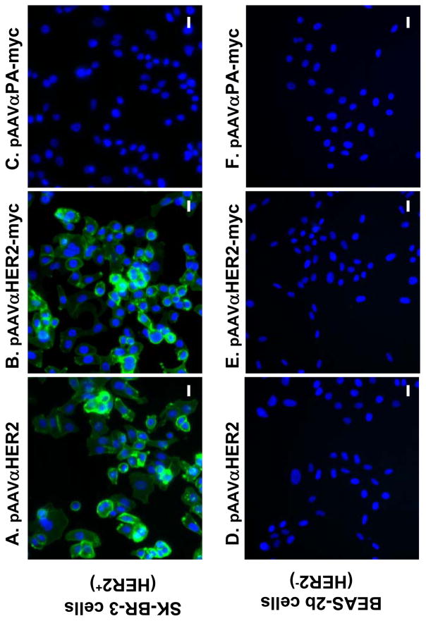
Specific binding of expressed anti-HER2 antibody to HER2 positive cells. Medium were collected from 293 cells transfected with pAAVαHER2, pAAVαHER2-myc and pAAVαPA-myc, respectively and added to fixed HER2 positive SK-BR-3 cells or fixed HER2 negative BEAS-2b cells. HER2 specific binding was assessed by immunofluorescent microscopy with Alexa 488 labeled goat anti-mouse second antibody. A. SK-BR-3 cells incubated with αHER2; B. SK-BR-3 cells, αHER2-myc; C. SK-BR-3 cells, αPA-myc; D. BEAS-2b, αHER2; E. BEAS-2b cells, αHER2-myc; and F. BEAS-2b cells, αPA-myc. All panels, bar = 20 μm.

