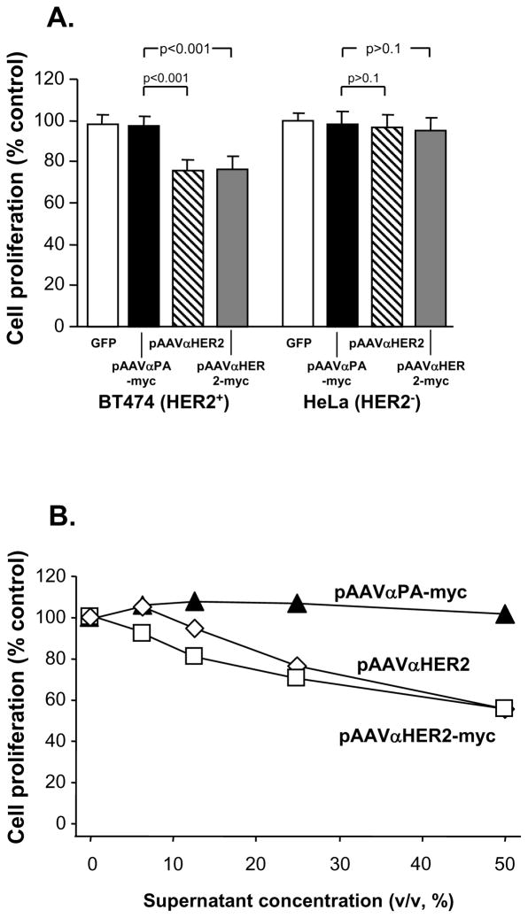
Figure 3.
Inhibition of HER2 positive cell proliferation by anti-HER2 antibody expressed by pAAVαHER2 and pAAVαHER2-myc plasmids. Media were collected from 293 cells transfected with mock, pGFP, pAAVαPA-myc, pAAVαHER2, pAAVαHER2-myc and added to HER2 positive BT474 cells and HER2 negative HeLa cells. After various time periods, cell proliferation was assessed. The data is expressed as the relative cell number compared to mock medium treated cells. Each point represents the mean of 8 replicates. A. Specific suppression of HER2+ cells proliferation by anti HER2 antibody expressed by the 293 cells. Medium from pGFP, pAAVαHER2, pAAVαHER2-myc or pAAVαPA-myc or transfected cells were added to BT474 (HER2+) or HeLa cells (HER2-) and cell proliferation assessed after 3 days. B. Dose-dependent suppression of HER2+ cells proliferation by anti HER2 antibody expressed by the 293 cells. Medium from pAAVαPA-myc, pAAVαHER2 or pAAVαHER2-myc transfected cells was 2-fold serial diluted and then added to BT474 cells. After 5 days, cells proliferation was assessed.

