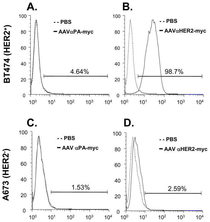
Figure 5.
HER2-specific cell binding from αHER2 antibody expressed in vivo following administration of AAVrh.10αHER2. PBS, AAVrh.10αHER2-myc or AAVrh.10αPA-myc (1011 ge-nome copies each) were administrated intravenously into C57Bl/6 mice and after 30 days serum was collected and pooled and used as the primary antibody. HER2 positive BT474 cells (panels A, B) and HER2 negative A673 cells (panels C, D) were exposed to the sera using FITC-labeled goat anti-mouse antibody and flow cytometry for detection. Solid lines - AAVrh.10αPA-myc sera or AAVrh.10αHER2-myc sera. Dashed lines - PBS control sera. A. BT474 cells exposed to sera from mice receiving the control vector AAVrh.10αPA-myc or PBS. B. BT474 cells exposed to sera from mice receiving AAVrh.10αHER2-myc or PBS. C. A673 cells exposed to sera from mice receiving the control vector AAVrh.10αPA-myc or PBS. D. A673 cells exposed to sera from mice receiving AAVrh.10αHER2-myc or PBS.

