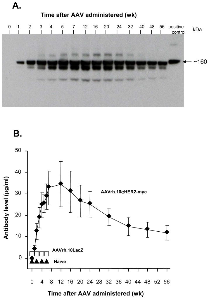
Figure 6.
Time course of AAVrh.10 mediated expression of anti-HER2 antibody in vivo. On day 0, C57Bl/6 mice (n=5/group) received intravenous administration of 1011 genome copies AAVrh.10αHER2-myc or AAVrh.10LacZ. Naive mice were used as an additional control. Serum was collected at different time points. A. Anti-HER2 antibody levels assessed by myc-specific Western analysis under non-reducing conditions. Serum from AAVrh.10αHER2-myc mice was pooled. Purified anti-HER2-myc was used as positive control. B. Serum anti-HER2 antibody levels were measured by human HER2-specific ELISA. Purified anti-HER2 antibody served as standard. Data was obtained from n=5 mice per group and is plotted as mean ± standard error.

