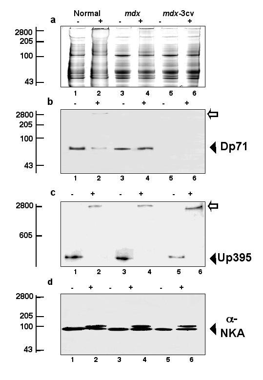
Figure 8.

Chemical crosslinking analysis of brain dystrophin isoform Dp71 in normal and mdx mice. Shown is a Coomassie-stained gel (a) and identical immunoblots labeled with antibodies to the dystrophin isoform of apparent 71 kDa (Dp71) (b), full-length utrophin of apparent 395 kDa (Up395) (c), and the α-subunit of the Na+/K+-ATPase (α-NKA) (d). Lanes 1, 3 and 5 represent untreated control samples (-) and lanes 2, 4 and 6 are membranes treated with 200 μg crosslinker BS3 per mg protein (+). Lanes 1 and 2, 3 and 4, and 5 and 6 represent microsomal membranes isolated from normal brain, mdx brain, and mdx-3cv brain, respectively. The position of immuno-decorated monomers is indicated by closed arrow heads and crosslinking-stabilized high-molecular-mass complexes marked by open arrows. The relative position of molecular mass standards (x 10-3) is indicated on the left.
