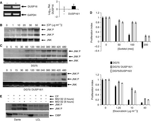
Figure 5.
DUSP16 expression modulates JNK activation and chemosensitivity in BL. (A) Ectopic expression of DUSP16 mRNA in DG75 cells transfected with DUSP16 expression vectors. DG75 cells were stably transfected with a DUSP16 expression plasmid as described in Materials and Methods section. DUSP16 mRNA levels were determined by RT–PCR (left panel) and qPCR (right panel) as described in Materials and Methods section. RT–PCR shows parental (−) and DUSP16-transfected (+) cell line analysed for DUSP16 and the control gene GAPDH. The qPCR shows the log10 of DUSP16 mRNA levels in one such clone of transfected cells (DUSP16/1) relative to parental DG75 cells (−)±1 s.d. (triplicate analyses). (B) Cisplatin induces dose-dependent activation of JNK in BL cells. Dante cells were exposed for 6 h to the indicated concentrations of cisplatin, after which cells were collected and total and phosphorylated JNK levels determined by western blotting as described in Materials and Methods section. JNK phosphorylation increases with increasing cisplatin concentration. (C) Ectopic expression of DUSP16 inhibits drug-induced activation of JNK. Parental DG75 cells and the clone DG75 DUSP16/1 were exposed to cisplatin (50 μg ml−1). Cells were collected at the indicated times (min) and the level of total and phosphorylated JNK determined by western blotting. Activation of JNK is detectable at 60 min in the parental cells, but not until 360 min in the cells ectopically expressing DUSP16. (D) Ectopic expression of DUSP16 reduces sensitivity of BL cells to cytotoxic drugs. Parental DG75 cells (black columns), together with two independent clones ectopically expressing DUSP16 (denoted DG75/DUSP16/1 (grey columns) and DG75/DUSP16/2 (white columns), were grown to logarithmic phase, then exposed to varying concentrations of sorbitol (mM) and doxorubicin (μg ml−1) as indicated. Proliferation was assessed using the CellTiter 96 Aqueous One solution cell proliferation assay (Promega) according to the manufacturer's instructions 48 h after addition of drug. Each point was determined in quadruplicate. Data shown are mean±1 s.d. (E) CtBP degradation in drug-treated cells is not influenced by JNK phosphorylation or DUSP16 status. Dante BL (in which DUSP16 is fully methylated) and LCLs were exposed to 50 μg ml−1 cisplatin for 6 h with or without MG132 for 3 or 6 h as shown, then subjected to western blotting analysis of phosphorylated JNK and CtBP. JNK phosphorylation occurs only in Dante cells, whereas CtBP degradation is seen in both cell lines and is unaffected by proteasome inhibition.

