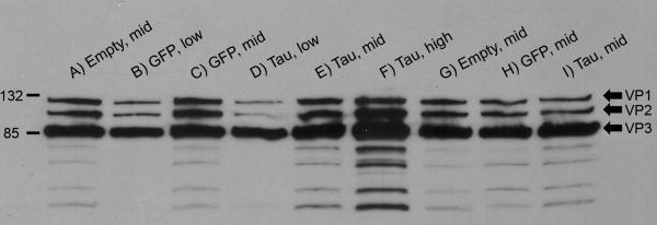
Fig 1.
AAV capsid protein western blot. Each viral dose used in the study was loaded and probed with an antibody against the AAV viral proteins VP1, VP2, and VP3 (indicated on right side of panel; size markers in kDa on left side), to ensure equal viral particles for the microarray study. Each matching dose of specific vectors had similar bands, and the intensity of the bands matched with the vector genome amounts loaded.

