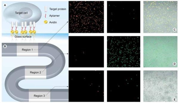
Figure 5. Microfluidic device and fluorescent images of captured cancer cells.
(A) Microfluidic device, (B) representation of aptamer immobilization, (C–E) enrichment of (C) CEM, (D) Ramos and (E) Toledo cells in their respective regions. Adapted with permission from [32] © American Chemical Society (2009).

