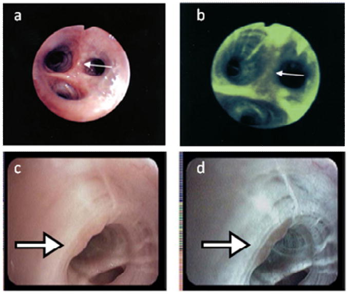
Figure 1.

These images illustrate the application of light induced fluorescence endoscopy to improve screening for lung cancer with a widefield optical imaging system; white light and autofluorescence images acquired with light-induced fluorescence endoscopy (top, Xillix LIFE; bottom, Pentax 3000) show typical image features associated with atypical cells, with suspicious areas indicated by white arrows (12, 13). The white light images (A, C) represent the clinician's conventional viewing mode, allowing anatomical inspection at low magnification. Autofluorescence images (B, D) are sensitive to the natural fluorescence of tissue, which originates predominantly from stromal collagen. The development of neoplasia is associated with loss of stromal autofluorescence and this loss of fluorescence intensity can be used to assist clinicians in identifying pre-cancerous and suspicious cancerous lesions (as shown by decreased fluorescence intensity within areas in B, D).
