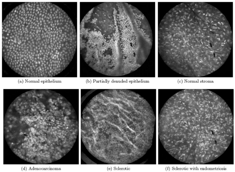
Figure 2.

High-resolution confocal microendoscope images of excised ovarian tissue after the application of acridine orange (16). Fluorescence imaging enabled visualization of individual nuclei and the underlying stroma (A-C). Images from a healthy ovary were characterized by a homogeneous distribution of nuclei, whereas images from carcinoma revealed a disordered tissue structure and variable nuclear size (A, D). Other more subtle pathologic changes such as sclerosis and endometriosis were also detectable (E, F).
