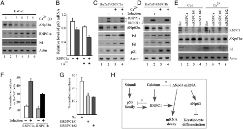
Fig. 5.
RNPC1 promotes keratinocyte differentiation by repressing p63 expression. (A) HaCaT cells grown at confluence were treated with or without 1.5 mM calcium for 0–9 d, and the level of ΔNp63α, RNPC1a, Ivl, and actin was measured by Western blot analysis. (B) The level of p63 transcript was measured in HaCaT cells uninduced or induced to express RNPC1a for 24 h along with or without treatment of 1.5 mM CaCl2 for 3 d. (C and D) The level of Involucrin and filaggrin along with ΔNp63, p21, and actin was measured in HaCaT cells treated as in (B). (E) The experiment was performed as in (C and D) with HaCaT cells transfected with scrambled siRNA or siRNAs against total RNPC1 or RNPC1a for 3 d, followed with or without treatment of CaCl2 for 3 d. (F) Confluent HaCaT cells were uninduced or induced to express RNPC1a or RNPC1b for 24 h, followed by treatment of 1.5 mM calcium for 9 d. Cornified cell envelopes were counted and expressed as percentage of total cells (mean ± S.D.; n = 3). (G) HaCaT cells were transfected with scramble siRNA or siRNA against RNPC1 for 3 d, followed by treatment of 1.5 mM calcium for 11 d. Cornified cell envelopes were counted and expressed as percentage of total cells (mean ± S.D.; n = 3). (H) A model of the RNPC1-p63 feedback loop and the role of RNPC1 in keratinocyte differentiation.

